APP Angle: ASGE APP Case of the Month

APP Angle. Resources for APPs.


Case 22: Upper GI Bleeding

A 56-year-old male with a history of alcoholic cirrhosis presents to the ER with new onset bright red bloody emesis. It began two hours ago. He had three episodes of bright red bloody emesis before arriving in the ER. The first episode was the “worst.” He vomited approximately 1 cup of bright red blood. The two subsequent episodes were smaller in volume, approximately one-fourth cup. He denies melena, bloody stools and abdominal pain. Two months ago, he had normal hemoglobin and a platelet count of 96,000. He has no prior history of GI bleeding and has not had a prior upper endoscopy for any indication. He denies alcohol use in the last three years. He does not use ASA or NSAIDs. He takes lisinopril for hypertension. In the ER, his CBC showed an Hgb of 10.2 and a platelet count of 92,000. His blood pressure was 98/64 with a heart rate of 98.

The most likely cause of his hematemesis is:

A. Duodenal ulcer
B. Esophageal varices
C. Gastric cancer
D. Gastric ulcer

The correct answer is B, esophageal varices. This case will only discuss acute esophageal variceal bleeding.

Practice Pearls

Natural History and Progression of Esophageal Varices

  • Approximately 50 percent of people with cirrhosis develop esophageal varices. The risk of bleeding (first episode) is 12 percent.1 Each episode of bleeding from esophageal varices is associated with up to 20 percent mortality.1
  • The formation of esophageal varices is a complication of portal hypertension. Cirrhosis is the most common cause of portal hypertension. Varices develop to decompress the hypertensive portal vein and return blood to the systemic circulation.1
  • The rate of progression of esophageal varices has not been well studied. A large prospective study noted that small esophageal varices progress in size at a rate of 12 percent at year one and 31 percent at year three.2
  • Predictive factors for esophageal variceal bleeding include1:
    • Location of varices (distal esophagus)
    • Size of varices (large varices)
    • Appearance of varices (red whale marks, cherry red spots)
    • Clinical features of the patient (advanced Child-Pugh classification, history of esophageal variceal bleeding)
    • Variceal pressure (if measured, bleeding risk begins at >13 mm Hg)

Clinical Manifestations

  • Symptoms can include anorexia, abdominal discomfort, jaundice, pruritis, altered mental status (encephalopathic symptoms) and muscle cramps.3
  • Signs of gastrointestinal bleeding, such as hematemesis, hematochezia (rapid gastrointestinal bleeding) and/or melena, due to esophageal varices can sometimes be the first clinical manifestation.3
  • Other physical examination findings associated with esophageal varices can include findings related to hypovolemia/anemia (hypotension, tachycardia) and features of portal hypertension (splenomegaly, ascites and caput-medusae).3

Management

  • It is important to ensure that the patient’s airway is protected, along with hemodynamic resuscitation. Prophylactic antibiotics, such as a fluoroquinolone or third-generation cephalosporin, should be given to patients with cirrhosis and upper GI bleeding.4
  • Pharmacologic therapy, such as terlipressin or octreotide, should be initiated in those with varices or at risk for varices, and should generally be continued for two to five days after bleeding cessation.4 Chelation therapy is reserved for patients who are intolerant or refractory to phlebotomy.2
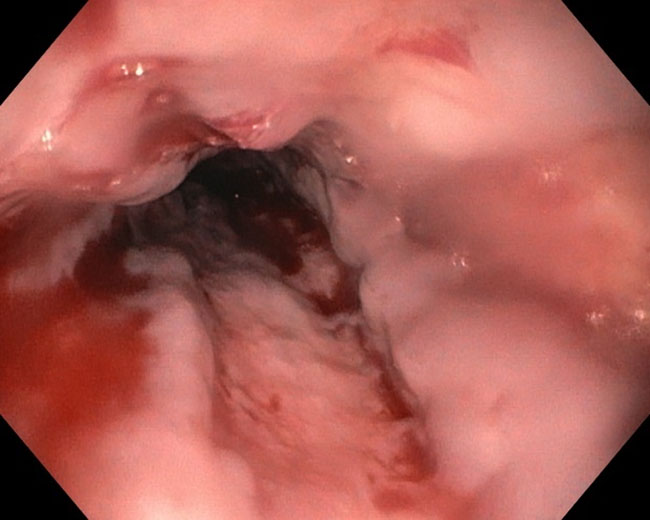
  • Endoscopic therapy is the treatment of choice for acute esophageal variceal hemorrhage (images 1 and 2) with the goal of EGD with endoscopic therapy within 12 hours of presentation.2,5
  • Endoscopic variceal ligation (EVL) is preferred as the initial treatment. 5 Elastic bands are placed on varices in the distal 5 cm of the esophagus.5 EVL has a success rate of approximately 90 percent.
  • Complications of EVL include worsening esophageal ulceration and development of portal hypertension in esophageal or gastric varices.5 Occasionally, patients may develop post-EVL chest discomfort.
  • Endoscopic sclerotherapy (injection of sclerosant into varices) is generally reserved for treatment failure with EVL.
  • In patients with rebleeding after achieving hemostasis with endoscopic therapy, a second EGD with endotherapy may be performed. If endoscopic therapy fails, transjugular intrahepatic portosystemic shunt (TIPS) should be considered. During TIPS, using a needle catheter, a stent is placed from the hepatic vein into the intrahepatic portion of the portal vein.5


Image 1
Bleeding Esophageal Varices
Images from the personal library of John A. Martin, MD, FASGE

Image 2
Bleeding Esophageal Varice
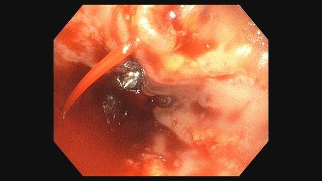
Images from the personal library of John A. Martin, MD, FASGE


References

  1. Sanyal AJ, Bajaj JS. Pathogenesis of variceal bleeding in patients with cirrhosis. UpToDate. Updated June 21, 2023. https://www.uptodate.com/contents/pathogenesis-of-variceal-bleeding-in-patients-with-cirrhosis
  2. Merli M, Nicolini G, Angeloni S, et al. Incidence and natural history of small esophageal varices in cirrhotic patients. J Hepatol. 2003;38:266-272.
  3. Meseeha M, Attia M. Esophageal varices. Stat Pearls. Published February 2019. Updated August 2, 2023. https://www.ncbi.nlm.nih.gov/books/NBK448078/
  4. De Franchis R, Burroughs A. After Groningen, other meetings followed. J Hepatol. 1990;63:743-752.
  5. Bajaj JS, Sanyal AJ. Methods to achieve hemostasis in patients with active variceal hemorrhage. UpToDate. Updated February 21, 2024. https://www.uptodate.com/contents/methods-to-achieve-hemostasis-in-patients-with-acute-variceal-hemorrhage

Authors

Palak Patel, PA-C, is a physician assistant in the Division of Gastroenterology at Northwestern Medicine in Chicago, IL.
Sarah Enslin, PA-C, is a physician assistant at the University of Rochester Medical Center in Rochester, NY with over 10 years of experience as a practicing PA in GI. Sarah serves on several national GI committees and is a former member of the ASGE Practice Operations Committee and a current member of the ASGE APP Committee.
Joseph Vicari, MD, FASGE, joined Rockford Gastroenterology in 1997 and has served as managing partner. He previously served as chair of the ASGE Practice Operations Committee, Councilor on the ASGE Governing Board and currently serves as co-chair of the ASGE APP Committee.